Summary
Epistasis, in which protein fitness changes in a non-additive way, is rare in natural evolution and laboratory evolution. Simple statistical models with only additive effects can explain most (80-95%) of changes in activity between variants (1–2). This has been suggested by multiple studies, which found that the effect of mutations that improve Stability and thermostability was basically entirely additive (see figure below). (5–6) Park et al. were able to model 92-96% of variance in genome fitness by accounting exclusively for single-point and pairwise interactions plus a sigmoid nonlinearity; e.g., less than <5% of genomes in their test set showed third-order interactions. (7) That said, there are examples where linear models are unable to accurately model fitness ((2,1,8) with Spike protein/ACE2), so the type of statistical model still matters.
Details
Sarkisyan et al. found that some mutations killed fluorescence in some GFP backbones but not others, and that pre-existing evolutionary propensity (e.g., from a PSSM) could not predict this. (9) They conclude that this is the result of destabilization. this made an additive model of fluorescence inappropriate; from that perspective epistasis occurred in 30% of multi-substitution variants. They conclude that this is largely due to threshold robustness, and that these mutations are destabilizing. See Mutation memory half life. Anecdotally (8) came to a similar conclusion.
Beltran et al. measured >500k pathogenic variants in human diseases across 500 domains and found that additive models were sufficient to model almost all fitness changes. (3)
Escobedo et al. found that the effects of substitutions in the hydrophobic core of proteins could be explained by linear models. (6)
Alcantar et al. carried out a “combinatorially complete” analysis of mutations introduced during affinity maturation of de novo designed minibinders and found that the binding improvements were basically entirely additive, which is similar to antibodies (Mutations obtained by antibodies during affinity maturation show epistasis in biophysical properties but not binding). (4)
Figures
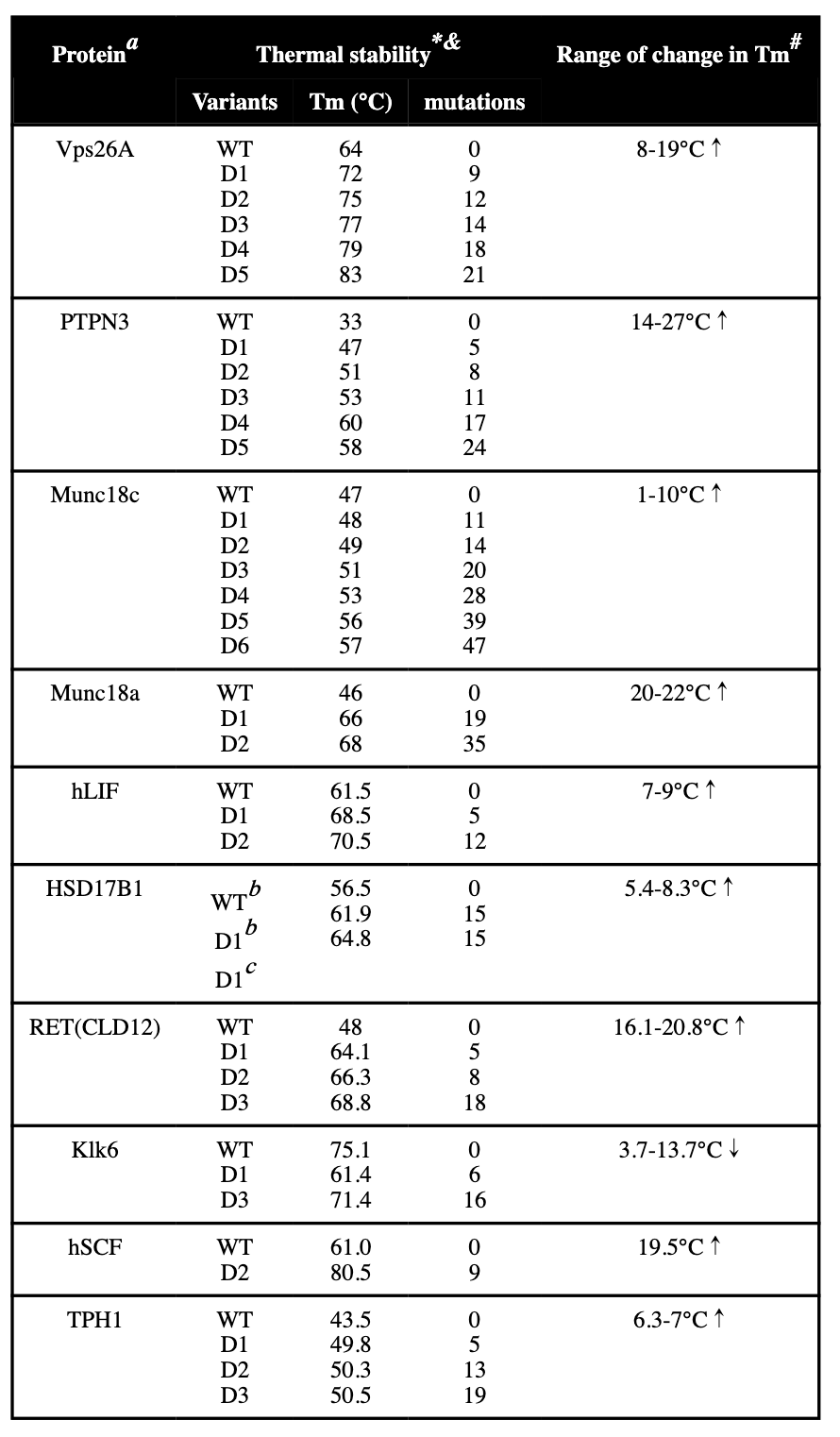 Ref (5)
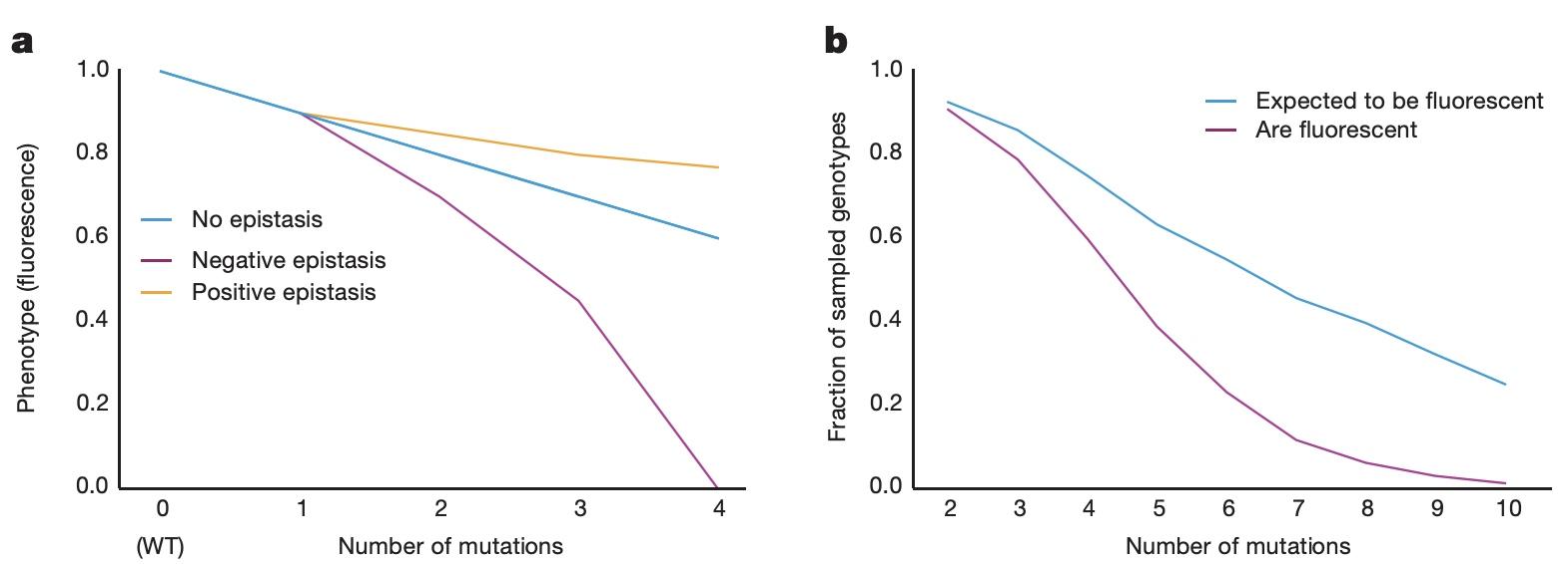 Ref (9)
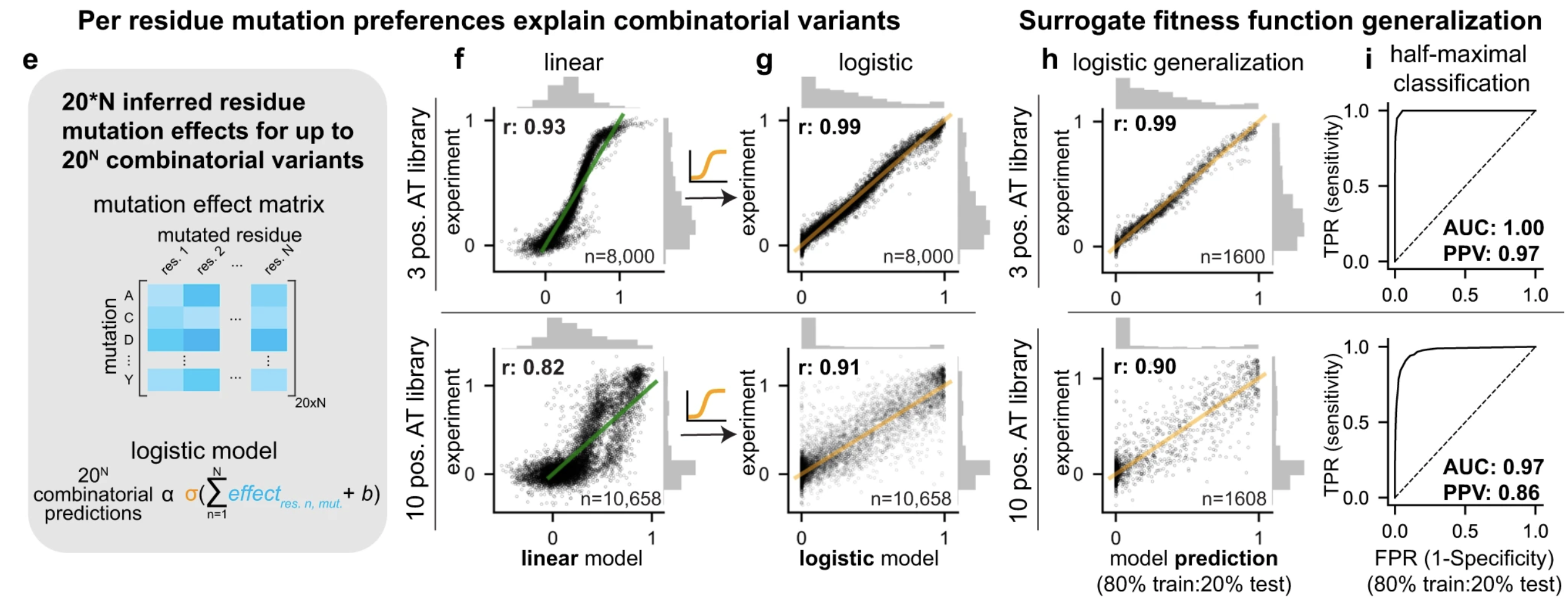 Figure 2 from (1)
See also
- Stability-activity trade-off during enzyme design and evolution is highly local and not global
- Mutations obtained by antibodies during affinity maturation show epistasis in biophysical properties but not binding